


In a major medical science breakthrough, scientists have developed the world’s first synthetic embryo outside the womb using stem cells cultured in a petri dish. These cells were cultured without the use of fertilized eggs, bypassing the need for sperm.
What is this synthetic embryo?
Researchers at the Weizmann Institute in Israel discovered that it was possible to get mouse stem cells to self-assemble into structures that resembled early embryos and had an intestinal tract, a developing brain, and a beating heart. These live constructs are often referred to as synthetic embryos because they are made without fertilized eggs. Moreover, they are anticipated to advance our understanding of how organs and tissues develop throughout the growth of natural embryos in the near future.
The study offers hopes of growing tissues and organs for transplantation using synthetic embryo models. The team tried to emulate what an embryo does and built on two previous advances which include an efficient method for reprogramming stem cells back to a naïve state and using an electronically controlled device that keeps the embryos bathed in a nutrient solution inside of beakers that move continuously, simulating the way nutrients are supplied by material blood flow to the placenta.
The team grew the embryo from mouse stem cells
The study was published in the journal Cell. It highlights the potential of naïve pluripotent cells to self-organize and functionally reconstitute. These cells can model the entire mammalian embryo. The team grew the embryo from mouse stem cells that had been cultured for years in a petri dish.
“Until now, in most studies, the specialized cells were often either hard to produce or aberrant, and they tended to form a mishmash instead of well-structured tissue suitable for transplantation. We managed to overcome these hurdles by unleashing the self-organization potential encoded in the stem cells,” Prof. Jacob Hanna of Weizmann’s Molecular Genetics Department, who headed the research team said.
The Process
The researchers separated the stem cells into three groups, one contained cells intended to develop into embryonic organs themselves, and the other two groups were pretreated for only 48 hours to overexpress one of two types of genes: master regulators of either the placenta or the yolk sac.
Thereafter, they were then mixed in the electronically controlled device, in which just about 0.5% or 50 of around 10,000 went on to form spheres. Each of which later became an elongated, embryo-like structure. They were able to observe the placenta and yolk sacs forming outside the embryos. Further, the model’s development proceeds as in a natural embryo.
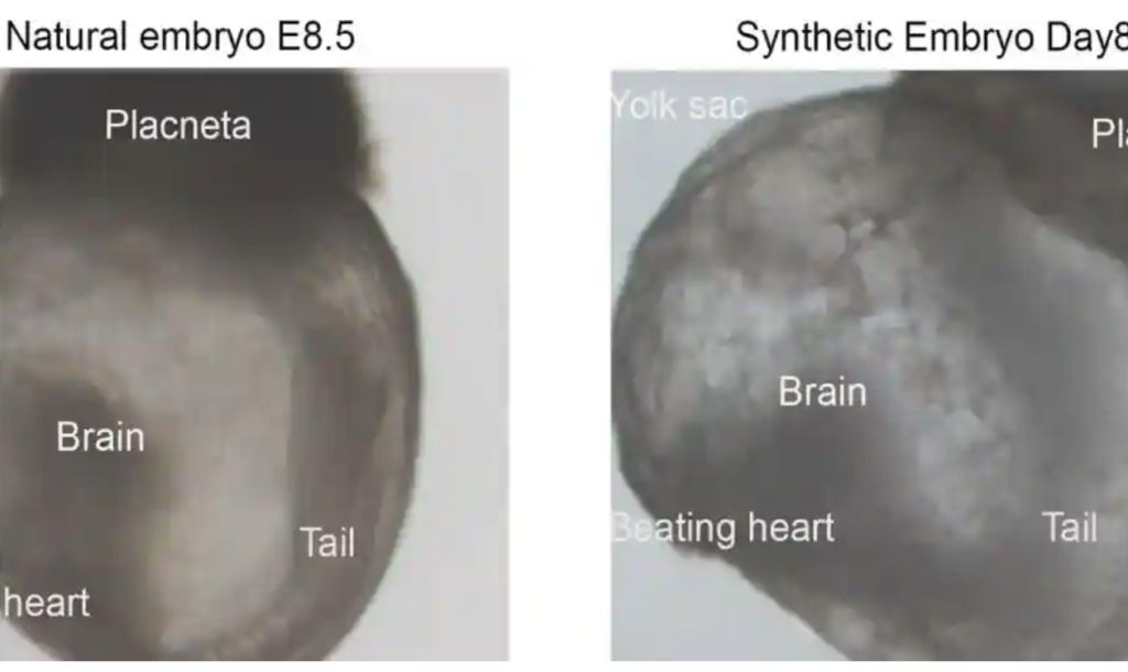
The synthetic embryos continued to develop for 8.5 days nearly half of the mouse’s 20-day gestation at which stage all the early organ progenitors had formed, including a beating heart, blood stem cell circulation, and a brain with well-shaped folds, a neural tube, and an intestinal tract.
When compared to natural mouse embryos, the synthetic models displayed a 95 percent similarity
“When compared to natural mouse embryos, the synthetic models displayed a 95 percent similarity in both the shape of internal structures and the gene expression patterns of different cell types. The organs seen in the models gave every indication of being functional,” researchers said in a statement.
The team is now looking to understand how stem cells know what to do. They are researching how they self-assemble into organs and find their way to their assigned spots inside an embryo. “Because our system, unlike a womb, is transparent, it may prove useful for modeling birth and implantation defects of human embryos,” Hanna added.